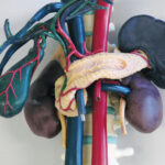
Anatomical Model for Ultrasound education
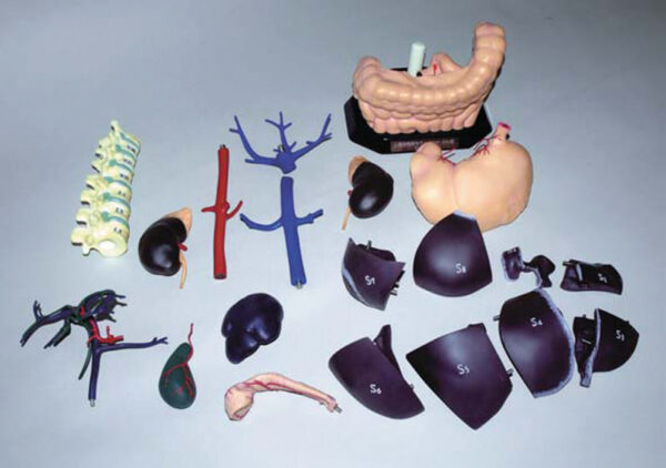
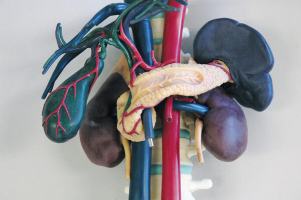
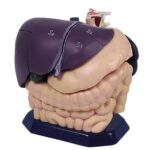
This 20 part model of the upper abdominal organs represents exactly the anatomy that is inside the training models R16560 and R16570. This allows you to see the structures and organs three dimensional in front of you while you are scanning them in the training model. The single parts are: liver (can be separated into 8 segments), gall bladder, spleen, left kidney, vena cava, spine, large and small intestine, portal
vein, bile duct and hepatic artery, pancreas, right kidney, abdominal aorta, hepatic vein and stomach.
vein, bile duct and hepatic artery, pancreas, right kidney, abdominal aorta, hepatic vein and stomach.